Brief:
Endoscopic Ultrasound (EUS) in Diagnosis and staging of pancreatic cancer
Description:
A 52 yrs old male patient was referred to us for the favour of EUS to evaluate the exact status of pancreas in view of recent episodes of acute pancreatitis. Patient has lost 5 kgs in the last few weeks and is complaining of recurrent pain off and on. All other imaging studies have been inconclusive so far.
-
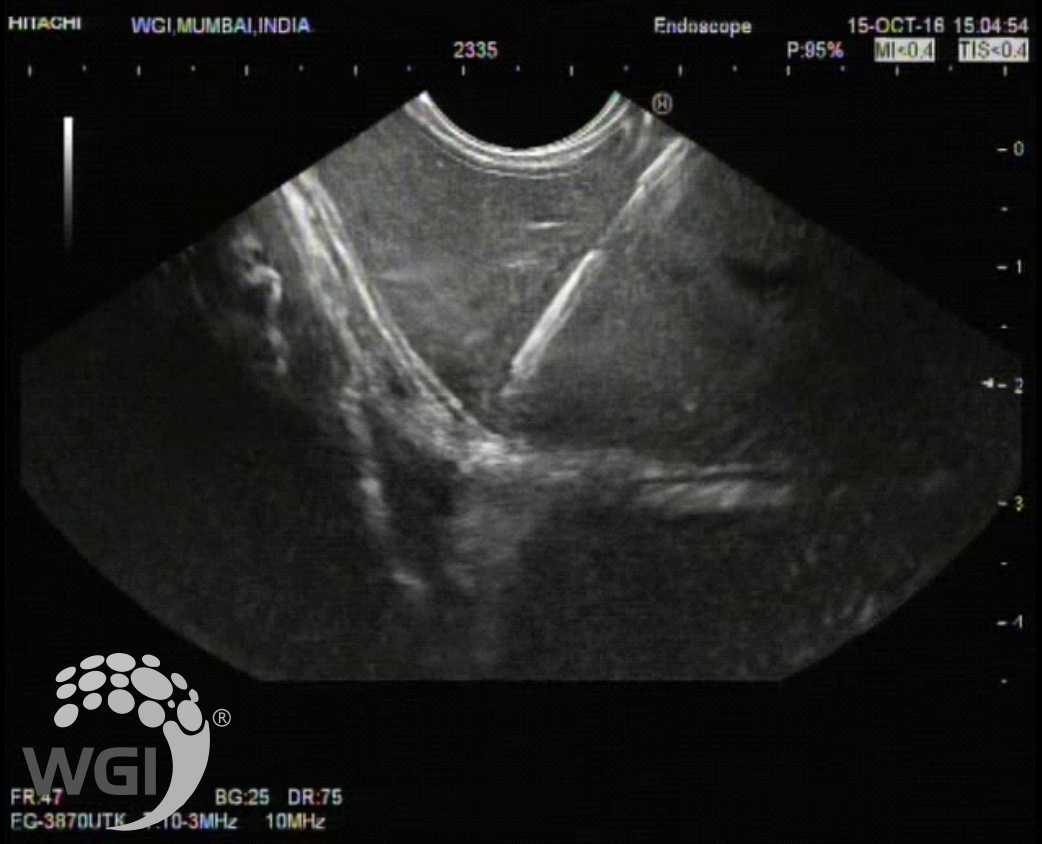
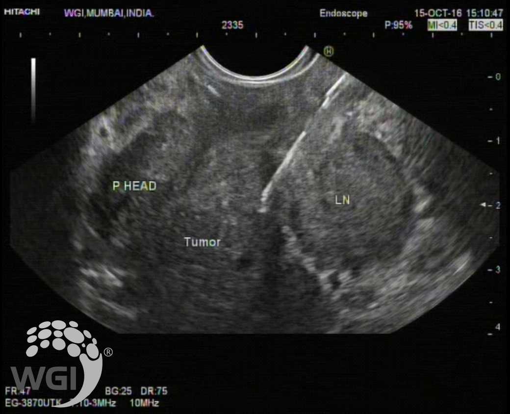
EUS revealed a large hypoechoic mass lesion in the pancreatic head region ( 3.5 cms x 3.0 cms) with multiple large nodes.
-
The mass appeared hard on Elastography. Contrast enhanced EUS was then performed which revealed hypoenhancement in the lesion suggestive of neoplastic process.
-
A lesion was also noted in the left lobe of liver which measured around 11 mm x 14 mm.
-
In view of these findings EUS guided FNA was then performed first from the left lobe liver lesion under color doppler control with a 22 g needle.
-
This was followed by one pass into the primary mass in the pancreatic head region. Aspirated material was sent for cytological examination and cell block preparation.
TAKE HOME MESSAGE
As we have seen in this case that whenever you suspect a pathology in Pancreas with the clinical history or biochemical abnormalities suggestive of pancreatic disease along with USG abdomen / CT Scan, the most important imaging modality that will provide specific additional information regarding the pathology is EUS. Here were able to diagnose the case of pancreatic adenocarcinoma based on EUS FNA with metastatic liver lesion. To support EUS imaging, we have more advanced features such as Contrast Enhanced EUS and Elastography to guide our targeting for the FNAB. Hence, in all the leading HPB centres, CT guided biopsies for pancreatic pathology is not preferred anymore and same is our experience in our unit for the last 19 years. We hope that clinicians would embrace this world class protocol in their practice and help the patient with accurate diagnosis and most optimal treatment in cases of pancreatic diseases.
Image:
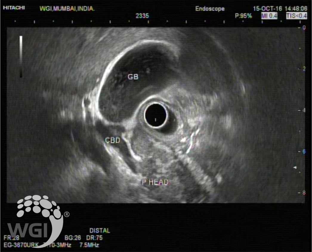
1. A large hypoechoic mass lesion in the pancreatic head region
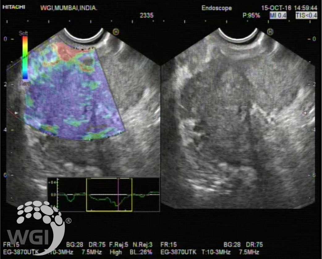
2. The mass appeared hard on Elastography
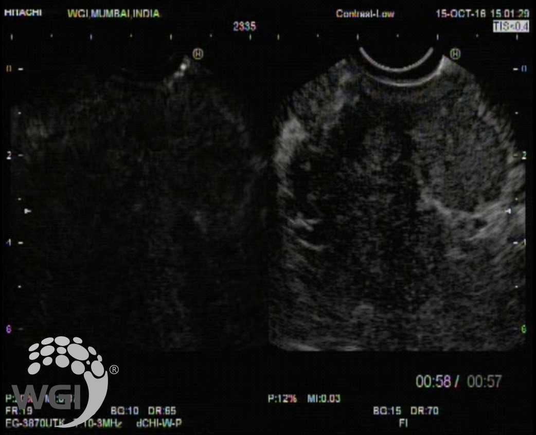
3. Contrast enhanced EUS was then performed
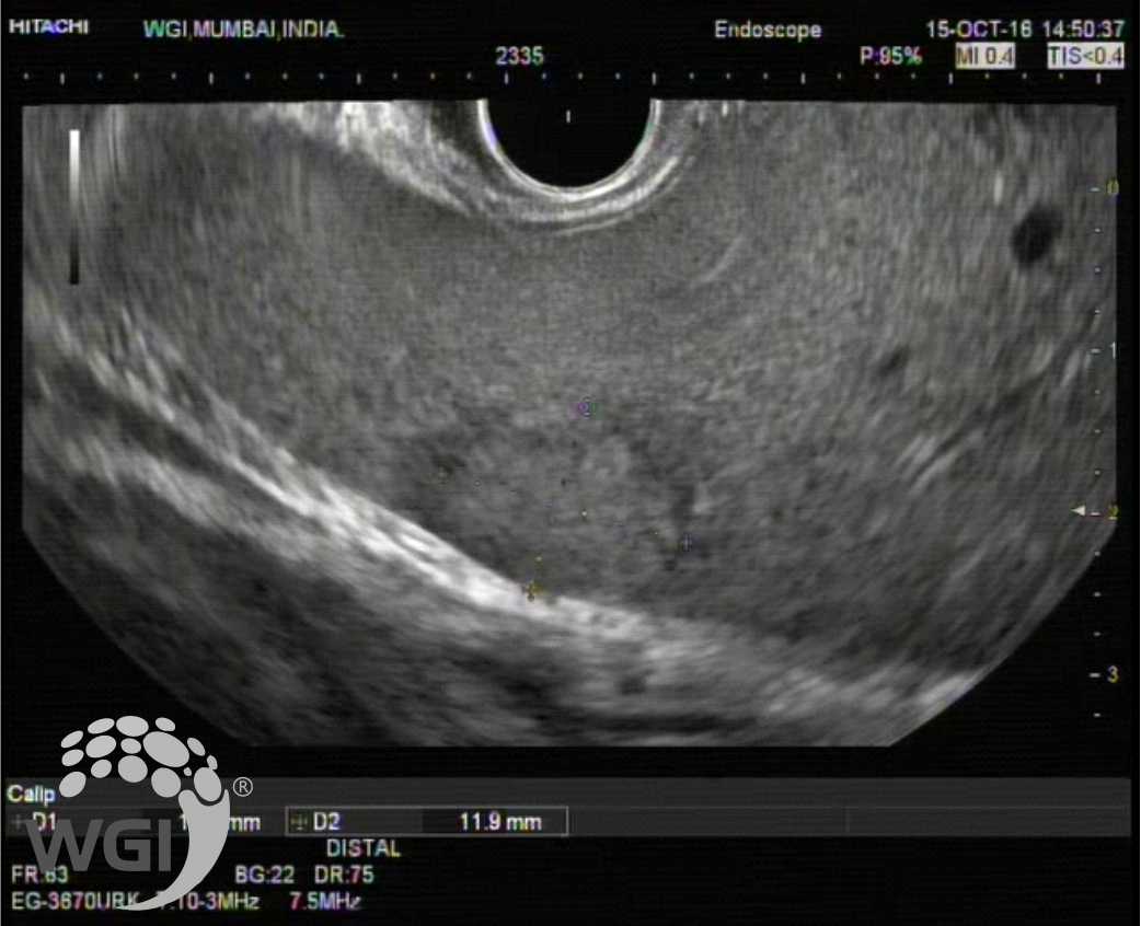
4. A lesion was also noted in the left lobe of liver
5. EUS guided FNA was then performed first from the left lobe liver lesion
6. EUS guided FNA was then performed from the pancreatic head region.
Posted by Dr. Vipulroy Rathod
Feb 27, 2019
Categories:
Gastrovision Case Capsules
© Endoscopy Asia