Brief:
ENDOSCOPIC ULTRASOUND DIAGNOSIS, AND PALLIATION OF MALIGNANT OBSTRUCTIVE JAUNDICE- EUS GUIDED CHOLEDOCHODUODENOSTOMY
Description:
-
A 55 yrs old female to referred to us for the favor of ERC and metal biliary stenting for an advanced metastatic pancreatic cancer invading the duodenum leading to cholestatic symptoms.
-
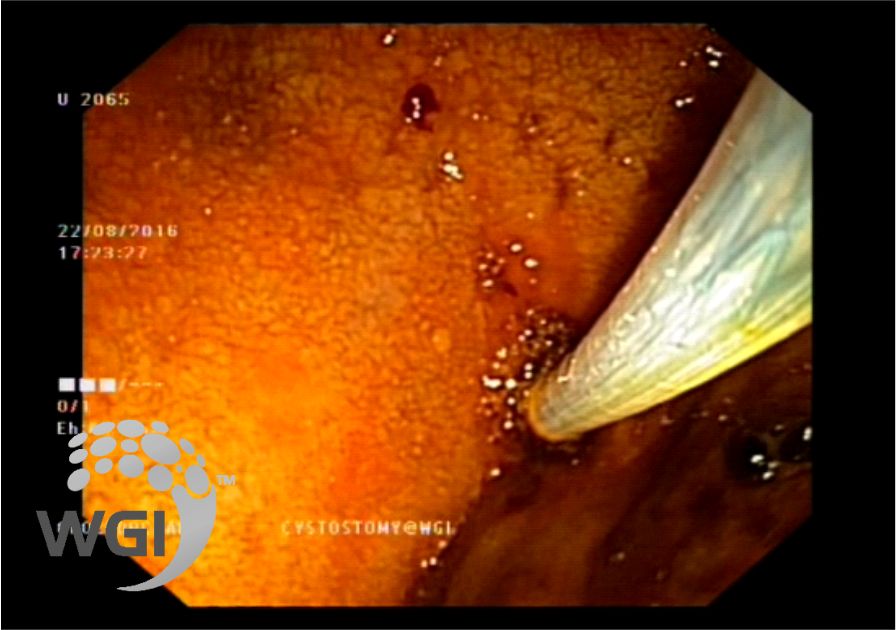
Duodenoscopy revealed large ulcerating mass in the second part of duodenum extending upto the D3.
-
The ampullary anatomy was completely distorted and despite several attempts selective cannulation of the CBD
-
EUS was then performed which revealed a massively dilated CBD with a block at the lower end on color doppler and EUS guided punctured of the CBD
-
A guidewire was placed and sequential dilatation of the CD
-
A 4 cms completely covered Metal stent was then deployed on Endoscopy and Fluoroscopy
TAKE HOME MESSAGE
As you have seen in this case that when conventional ERCP is not possible from the transpapillary route and when even PTC is not feasible due to extensive tumor and minimal ascites, EUS guided biliary drainage procedure proves to be of great value. I remember I was fortunate to pioneer this procedure-EUS Guided Choledocho-duodenostomy for the first time in India and even Asia way back in 2001. Now we hear various centres talking about EUS guided Biliary Drainage. However, a word of caution, this procedure should be performed only by those who have extensive interventional EUS experience and expertise. Furthermore, careful case selection is of paramount importance to avoid and minimize adverse events.
Image:
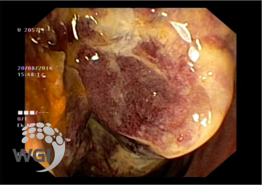
1. Large ulcerating mass in the second part of duodenum extending upto the D3
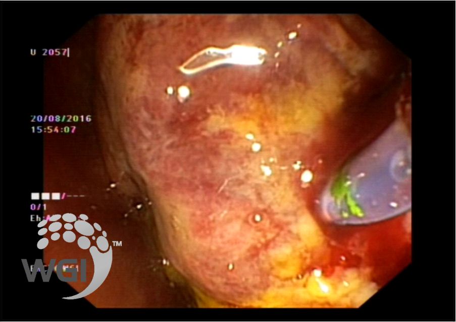
2. Attempts at selective cannulation of the CBD transpapillary failed
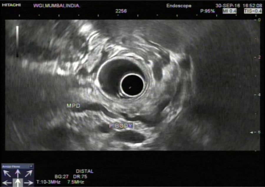
3. A massively dilated CBD with a block at the lower end on color doppler
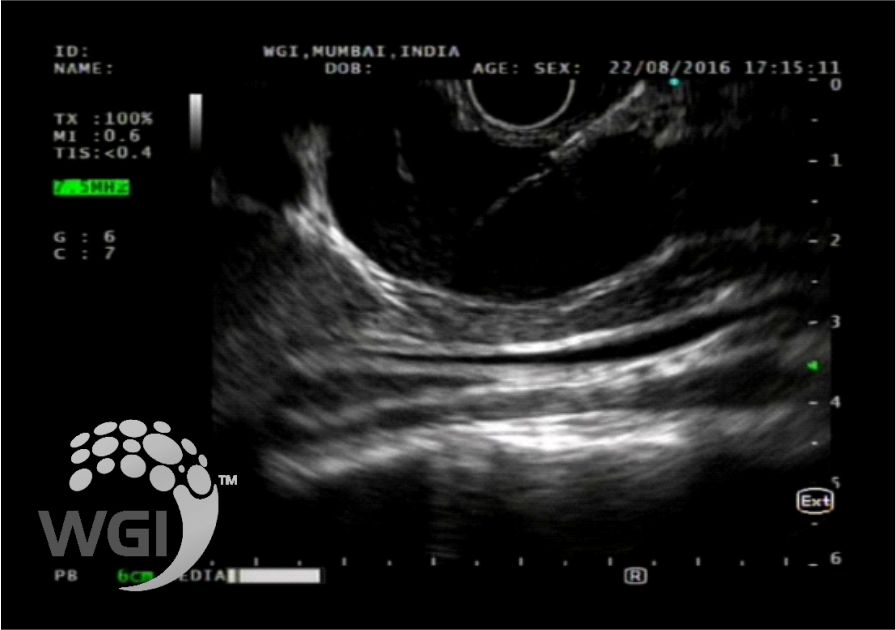
4.EUS GUIDED puncture of the CBD
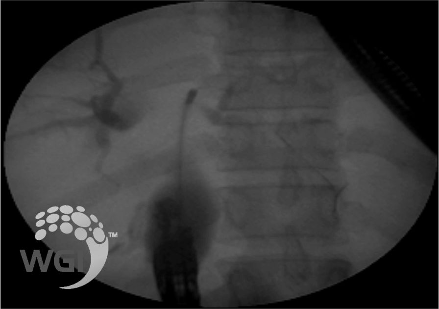
5.A guidewire was placed and sequential dilatation of the CD
6.Fully covered metal stent passed through the duodenal bulb
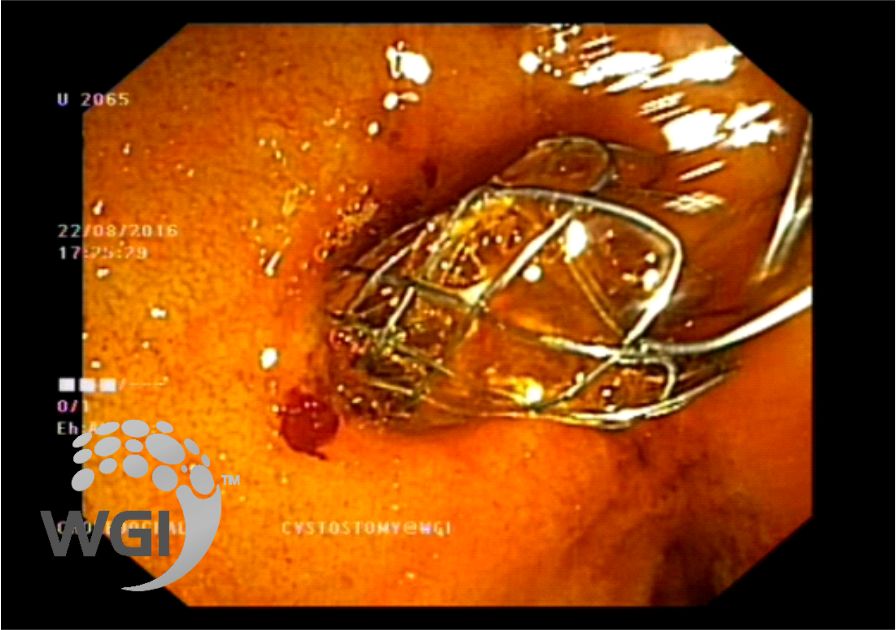
7.A 4 cms completely covered Metal stent was then deployed and free flow of dark bile was seen.
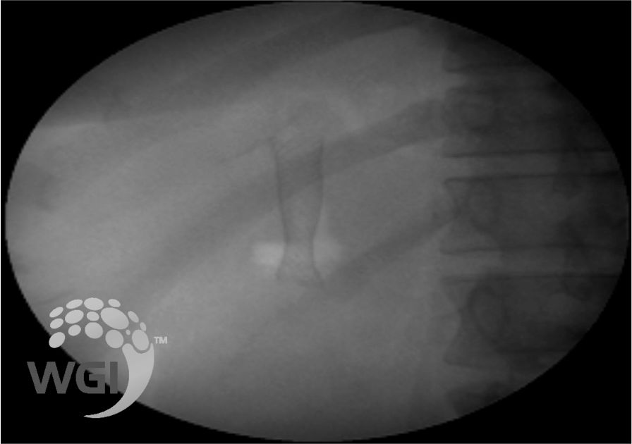
8.Fluoroscopy showed a 4 cms covered metal stent was deployed
Posted by Dr. Vipulroy Rathod
Dec 05, 2016
Categories:
Gastrovision Case Capsules
© Endoscopy Asia